The Cancer Foundation of Santa Barbara is pleased to support the design, construction and installation of this new digital technology for the Santa Barbara community.
What is PET/CT Imaging?
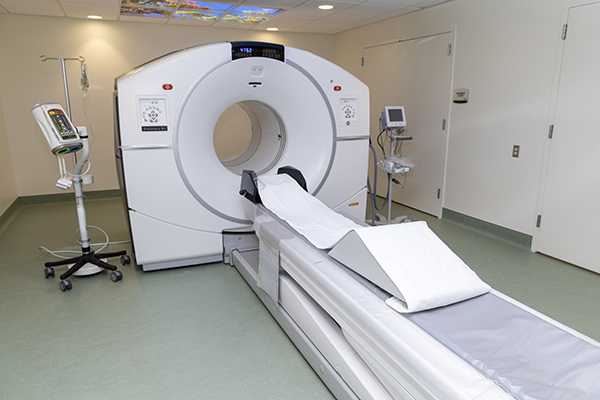
Positron Emission Tomography (PET) scans use radioactively labeled tracers (radiotracers) that are injected into the bloodstream. Radiotracers are small molecules that are designed to be very similar to compounds normally found in the body. These specially marked compounds emit positrons as they break down. PET scanners contain special cameras that detect these positrons and create images that show how the cells are functioning within the body. The image distinguishes areas that have little radiotracer concentration from areas with increased concentrations of radiotracer. On an image, the differences are represented by color changes.
A Computed Tomography (CT) scan creates detailed images of the structure of organs in the body. The scanner rotates around the patient while shooting X-ray beams toward them. A computer that is connected to the machine then creates separate slices, or images, of the body. A doctor can thread the slices together to examine the different structures of the body.
By combining the power of each of these machines into one PET/CT scanner, the nuclear medicine physician receives information regarding both the structure and function of the cells and organs in the body in one sitting. It is important to have molecular information in addition to structural information in the oncology realm because tumors can often leave scarring after therapy so CT imaging alone isn’t as accurate in determining how the cells have responded to therapy.
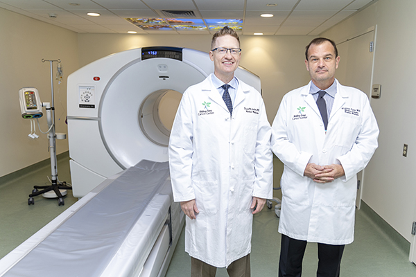
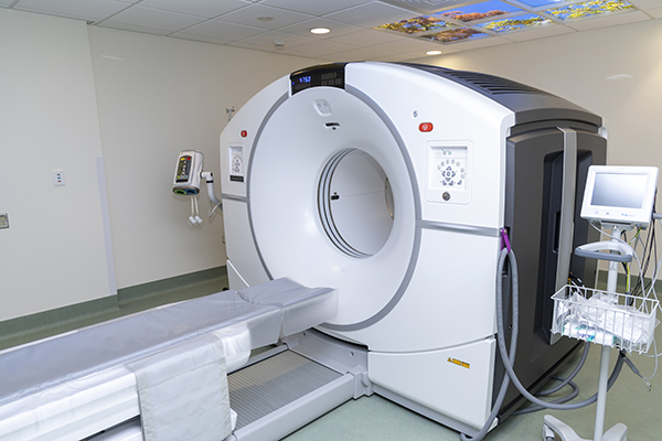
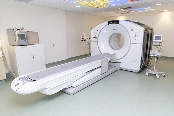
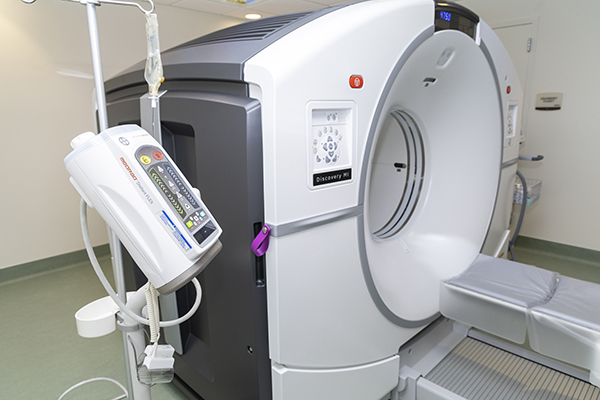


Digital PET/CT Offers Many Benefits
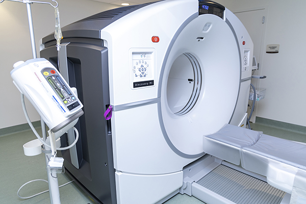
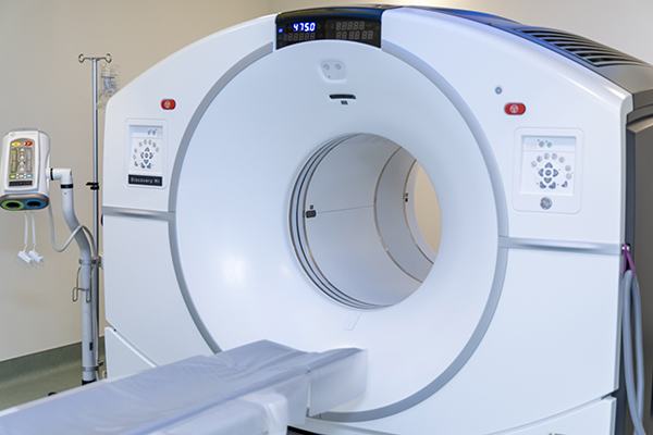
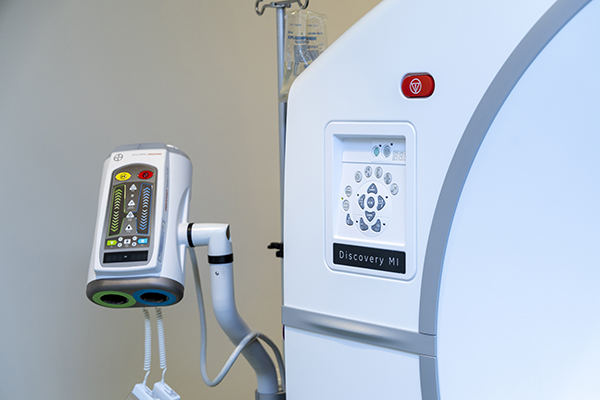
DECREASED RADIATION AND IMPROVED IMAGES – New digital PET/CT imaging technology improves image resolution by 2 times allowing for detection of smaller lesions, while reducing CT radiation dose by up to 82%.
IMPROVED PATIENT OUTCOMES – Digital PET/CT images show disease that would potentially be missed with a non-digital system. This enhancement increases diagnostic confidence, allows early diagnosis and potentially improves patient outcomes.
ENHANCED PATIENT EXPERIENCE – Nuclear medicine exams are notoriously long. The new scanners allow for images to be obtained at a much faster rate while providing exceptional image quality. This provides increased access to screening exams, and reduced wait time for exams that detect early onset of disease.
MOTION CORRECTION – More than 50% of patients should have a motion corrected PET/CT exam, but less than 5% of patients get one (breathing during imaging results in loss of information). GE’s Digital PET/CT provides motion correction for every patient, without the need for cumbersome devices. The high count rate of the digital PET allows for device less motion correction.
CUTTING-EDGE THERAPY – Ridley-Tree Cancer Center recently began PET/CT imaging for coronary artery disease, neuroendocrine tumors and prostate cancer. Digital PET/CT allows truly cutting-edge therapy evaluation with treatment radiopharmaceuticals such as Y-90 TheraSpheres.
To see if a PET/CT scan is important to your care, please call (805) 682-7300.
What is SPECT/CT Imaging?
SPECT/CT is an imaging process in which two different types of scans are taken and the images or pictures from each are fused or merged together. The fused scan can provide more precise information about how different parts of the body function and more clearly identify problems such as tumors (lumps) or Alzheimer’s disease, etc.
SINGLE PHOTON COMPUTED TOMOGRAPHY (SPECT) images are obtained following the administration of a small amount of radioactive material called a radiopharmaceutical that is used for nuclear medicine scans. This medication is taken up by cells and organs in the body depending on which radiopharmaceutical is used. For example, if cancer is suspected to have spread to the bones a radiopharmaceutical that mimics molecules that the bones use to repair themselves is injected into the patient. The radioactivity coming from the patient is then detected, and three-dimensional pictures are produced by the SPECT camera.
COMPUTED TOMOGRAPHY (CT) Images are obtained while the patient lies on a bed that moves into a ring, or “donut” shaped X-ray machine. The X-ray machine rotates over a 360 degree arc around the patient, allowing for image reconstruction in three dimensions. The CT scanner rotates much faster than the gamma camera, so the CT part of the study takes less time than the SPECT study.
The similarity between SPECT and CT in the method of image processing allows the images to be combined. Combining the information from a nuclear medicine SPECT study and a CT study allows the information about function from the nuclear medicine study to be easily combined with the information about how the body structure “looks” in the CT study.
Benefits of SPECT/CT
Due to direct photon conversion, the SPECT/CT offers improved image resolution and greater sensitivity. The energy resolution is 2x better and the improved flexibility of the camera allows for enhanced imaging. The table on the SPECT/CT can accommodate patients up to 500 pounds. Patients receive less radiation with a whole body scan, including three-dimensional imaging completed in 45 minutes compared to the current 75 minutes that are required.

